Non-Gynae Cytology Service
Liverpool Clinical Laboratories
Pathology

Contacts: Royal Liverpool University Hospital Non-gynae Cytology Laboratory
Tel: 0151 706 5623

Claire Chadwick, BMS2
Cytology Lab Manager
Tel: 0151 706 5623
Email: claire.chadwick@LiverpoolFT.nhs.uk

Robert Lee
Non-Gynae Cytology Team Leader
Tel: 0151 706 5623
Email: robert.lee@LiverpoolFT.nhs.uk

Location: Non-gynae Cytology Laboratory
Royal Liverpool Hospital
4th Floor CSSB
Vernon Street
Liverpool
L7 8YE

Opening Hours: Monday - Friday 09:00 - 17:30


Types of clinical services offered by the laboratory (including examinations referred to other laboratories)

The service is provided by Consultant Cytopathologists who undertake a full range of non-gynaecological cytology investigations, including respiratory specimens, urine, body cavity fluid, CSF, cyst fluid, and fine-needle aspiration cytology (FNAC) specimens. Synovial fluid is also examined for crystals, as part of the service to Orthopaedics and Rheumatology.

FNAC work is extensively used by the Breast Screening Programme. FNAC services are also provided to the Thyroid, Gastro and Regional Head & Neck Oncology Clinics.

Where patients are unable to attend the above clinics a similar rapid FNAC service is available to clinicians on the Royal Liverpool Hospital site for palpable lumps by individual request. Clinicians requesting this type of FNAC must contact Claire Chadwick, BMS2, giving at least 24 hours notice. Where possible, a provisional or final report will be issued within 1-2 hours.
Clinicians requesting this type of FNAC must contact Claire Chadwick, BMS2, giving at least 24 hours notice. Where possible, a provisional or final report will be issued within 1-2 hours.

The service also makes use of the Immunocytochemistry and Molecular Pathology facilities within the Department of Pathology.

Turnaround times

Turnaround time relates to the final local report. Turnaround times are defined from the time of collection from the patient to completion and confirmation of the test result so that it is available to the requestor.

In line with Royal College of Pathologist guidelines the Department aims for the following turnaround times:

90% of all requests reported final within 7 calendar days from date of collection.

98% of all requests reported final within 10 calendar days from date of collection.

URGENT results:

If a result is required urgently this request should be made directly to the laboratory (Tel. 0151 706 5623).

Instructions for completion of the request form and the laboratory’s criteria for accepting and rejecting samples

All tests must be commissioned using a request form which must accompany the sample. The appropriate Non-Gynae cytology request forms must be used to ensure that all the information required is included. All areas of the request form must be completed except for the section titled as ‘For Laboratory use only’. All relevant clinical information, including previous specimens submitted, previous tumours and any treatment given (such as radiotherapy or chemotherapy) should be recorded on the request form. The request form must be signed to show responsibility for the request. It is also important that the sample is labelled appropriately. Service users must ensure that they have checked all of the information on both the request form and sample, any discrepancies or missing information will lead to a delay in the sample preparation and reporting.

Service users must ensure that where samples are being sent to multiple disciplines a separate sample and request form are generated for each discipline required.

For details of the laboratory’s criteria for accepting and rejecting samples (Minimum Data Standard policy) click here .

Instructions for transportation of samples, including special handling needs

Samples must be transported to the laboratory as soon as possible to prevent deterioration of the cells. If transport is delayed then fresh samples should be refridgerated between 2-8C until transportation can be arranged. Samples collected in CytoLyt should be stored between 15-30C.

Examinations offered by the laboratory including, as appropriate, information concerning samples required, primary sample volumes, special precautions, turnaround time, biological reference intervals, and clinical decision values

BRONCHIAL WASHINGS, BRONCHOALVEOLAR LAVAGE (BAL)

Investigation Non-Gynae Cytology
Inform lab before sending No (unless very urgent: extension 5623)
Specimen type Bronchial Washings / BAL
These specimens are collected during bronchoscopy to investigate focal or diffuse lung abnormalities
Maximum Volume Required 25 mls
Specimen container
Sterile 30 ml universal
Make sure lid is firmly secured
Transport to the laboratory If transport is delayed then refrigerate sample. Delays of over 48 hours are undesirable
Turnaround 5 - 10 working days
Preparation method used by the laboratory ThinPrep Liquid Based Cytology.Papanicolaou Staining.
Factors known to significantly affect the performance of the examination or the interpretation of results The nature of the abnormality being investigated should be clear from the clinical information included with the specimen so that the laboratory can perform the appropriate preparations.

A negative result of an exfoliative cytology sample is not sufficient evidence to exclude significant disease. Discussion of cases at multi-disciplinary team meetings and submission of further samples, if deemed clinically appropriate, should thus be a routine part of the diagnostic pathway. Cytology results should be correlated with histology findings. Audit against final outcomes (which may be clinical) should be performed.
Additional information Please ensure that all request forms and specimen pots are clearly labelled with patient details and relevant clinical information.
Use a Non-Gynae request form/ICE request form


BRONCHIAL BRUSHINGS

Investigation Non-Gynae Cytology
Inform lab before sending No (unless very urgent: extension 5623)
Specimen type Bronchial brushing
Bronchial brushing are taken at bronchoscopy for the investigation of suspected tumours
Maximum Volume Required 20 mls
Specimen container
30 ml universal containing 10 mls of Cytolyt fluid.

These are obtained from Non-Gynae Cytology ext 5623.

The staff will send you a bag containing 20 bottles

Place uncovered brush tip into the universal and shake vigorously for a few seconds. LEAVE brush tip in universal.

All bottles have an expiry date printed on them.

Make sure lid is firmly secured
Transport to the laboratory If transport is delayed specimen will keep for about 1 month.
Turnaround 5 - 10 working days
Preparation method used by the laboratory ThinPrep Liquid Based Cytology. The literature indicates that better results are achieved with this approach than with direct smears prepared at the bedside.
Papanicolaou Staining.
Factors known to significantly affect the performance of the examination or the interpretation of results The nature of the abnormality being investigated should be clear from the clinical information included with the specimen so that the laboratory can perform the appropriate preparations.

The highest diagnostic yield is found when a visible abnormality is sampled.

Bleeding induced by biopsy of the lesion may obscure the cellular material and thus the brushing should be performed before a biopsy is taken.

If the brush tip is not exposed when placed in CytoLyt then the CytoLyt cannot penetrate the cells and this therefore affects the cellular preservation of the sample.

A negative result of an exfoliative cytology sample is not sufficient evidence to exclude significant disease. Discussion of cases at multi-disciplinary team meetings and submission of further samples, if deemed clinically appropriate, should thus be a routine part of the diagnostic pathway. Cytology results should be correlated with histology findings. Audit against final outcomes (which may be clinical) should be performed.
Additional information Please ensure that all request forms and specimen pots are clearly labelled with patient details and relevant clinical information.
Use a Non-Gynae request form/ICE request form


BILE DUCT BRUSHINGS

1. Bile duct brushings are used for the investigation of suspected neoplastic strictures of the biliary tree

2. They should be taken before therapeutic interventions are performed. Bile duct brushings should not be taken if a stent is in place as the reactive changes induced in the glandular epitelial cells will make the result unreliable.

Investigation Non-Gynae Cytology
Inform lab before sending No (unless very urgent: extension 5623)
Specimen type See above
Maximum Volume Required 20 mls
Specimen container
30 ml universal containing 10 mls of Cytolyt fluid.

These are obtained from Non-Gynae Cytology ext 5623.

The staff will send you a bag containing 20 bottles

Place uncovered brush tip into the universal and shake vigorously for a few seconds. LEAVE brush tip in universal.

All bottles have an expiry date printed on them.

Make sure lid is firmly secured

Transport to the laboratory If transport is delayed specimen will keep for about 1 month.
Turnaround 5 - 10 working days
Preparation method used by the laboratory ThinPrep Liquid Based Cytology. The literature indicates that better results are achieved with this approach than with direct smears prepared at the bedside.
Papanicolaou Staining.
Factors known to significantly affect the performance of the examination or the interpretation of results The nature of the abnormality being investigated should be clear from the clinical information included with the specimen so that the laboratory can perform the appropriate preparations.

If the brush tip is not exposed when placed in CytoLyt then the CytoLyt cannot penetrate the cells and this therefore affects the cellular preservation of the sample.

A negative result of an exfoliative cytology sample is not sufficient evidence to exclude significant disease. Discussion of cases at multi-disciplinary team meetings and submission of further samples, if deemed clinically appropriate, should thus be a routine part of the diagnostic pathway. Cytology results should be correlated with histology findings. Audit against final outcomes (which may be clinical) should be performed.
Additional information Please ensure that all request forms and specimen pots are clearly labelled with patient details and relevant clinical information.
Use a Non-Gynae request form/ICE request form


EBUS

Investigation Non-Gynae Cytology
Inform lab before sending No (unless very urgent: extension 5623)
Specimen type EBUS
Endobronchial ultrasound-guided transbronchial needle aspiration (EBUS) for mediastinal masses is performed to investigate mediastinal masses, predominantly in the context of staging of non-small cell lung cancer. Other conditions associated with mediastinal lymphadenopathy include cancer of other organs, atypical infections and sarcoidosis.
Maximum Volume Required 20 mls: When possible place EBUS sample from same site into same universal pot.
Specimen container
30 ml universal containing 10 mls of Cytolyt fluid.

These are obtained from Non-Gynae Cytology ext 5623.

All bottles have an expiry date printed on them.

Make sure lid is firmly secured

Transport to the laboratory Send to laboratory A.S.A.P. The sample will keep for 1 month if specimen delayed for any reason.
Turnaround 5 - 10 working days
Preparation method used by the laboratory ThinPrep Liquid Based Cytology, Papanicolaou Staining.
Cell block, Haematoxylin and Eosin staining. Immunocytochemistry is also required for the majority of cases.
Factors known to significantly affect the performance of the examination or the interpretation of results The nature of the abnormality being investigated should be clear from the clinical information included with the specimen so that the laboratory can perform the appropriate preparations.

A negative result of an exfoliative cytology sample is not sufficient evidence to exclude significant disease. Discussion of cases at multi-disciplinary team meetings and submission of further samples, if deemed clinically appropriate, should thus be a routine part of the diagnostic pathway. Cytology results should be correlated with histology findings. Audit against final outcomes (which may be clinical) should be performed.
Additional information Please ensure that all request forms and specimen pots are clearly labelled with patient details and relevant clinical information, including date/time sample taken.
Use a Non-Gynae request form or ICE generated form.


SPUTUM

Investigation Non-Gynae Cytology
Inform lab before sending No (unless very urgent: extension 5623)
Specimen type SPUTUM
This is recognised to be a specimen of limited or no clinical value, and hence should be rarely received.
Where patients are unfit for bronchoscopy and the patient has suspected lung cancer, three separate sputum samples collected on different days should be sent for cytological examination.
Nebulised saline may be used to induce sputum production in appropriate clinical circumstances.
Guidance should be given to the patient on producing a deep cough sample. A salivary sample is inadequate for cytology.
The whole of the expectorated sample should be submitted.
Maximum Volume Required Any sample provided
Specimen container
Sterile leak proof container.
Make sure lid is firmly secured
Transport to the laboratory Induced sputum specimens should reach the laboratory in a timely fashion. If transport is delayed then refrigerate sample. Delays of over 48 hours are undesirable
Turnaround 5 - 10 working days
Preparation method used by the laboratory ThinPrep Liquid Based Cytology, Papanicolaou staining.
Factors known to significantly affect the performance of the examination or the interpretation of results Sputum cytology should not be requested routinely in patients presenting with respiratory infections.

Sputum is not an effective investigation to screen for occult malignancy.

The nature of the abnormality being investigated should be clear from the clinical information included with the specimen so that the laboratory can perform the appropriate preparations.

If the patient has provided saliva or if there are visible food particles, it is better to start again straight away. Better samples may be forthcoming early in the morning (but before breakfast!), or with the assistance of a physiotherapist.

The quality of sputum deteriorates rapidly. Day-old specimens seldom provide any useful result.

If a sample requires microbiological tests, then a separate sample should be provided.

A negative result of an exfoliative cytology sample is not sufficient evidence to exclude significant disease. Discussion of cases at multi-disciplinary team meetings and submission of further samples, if deemed clinically appropriate, should thus be a routine part of the diagnostic pathway. Cytology results should be correlated with histology findings. Audit against final outcomes (which may be clinical) should be performed.
Additional information Please ensure that all request forms and specimen pots are clearly labelled with patient details and relevant clinical information.
Use a Non-Gynae request form/ICE request form


URINE

Investigation Non-Gynae Cytology
Inform lab before sending No (unless very urgent: extension 5623)
Specimen type URINE
Freely voided, catheter, ileal conduit specimens or bladder/ureteric washings may be collected. It is essential that the specimen collection method is documented on the request form. The first urine passed in the morning should be avoided. A mid-stream specimen is sub-optimal.
Samples may be taken from the upper tract by clinicians experienced in the technique and should be handled in the same way as urine specimens.
Maximum Volume Required A maximum of 20 ml of fresh sample is required. For voided urine, an aliquot of the whole voided sample should be submitted.
Specimen container
Sterile 30 ml universal
Make sure lid is firmly secured
Transport to the laboratory If transport is delayed then refrigerate sample. Delays of over 48 hours are undesirable
Turnaround 5 - 10 working days
Preparation method used by the laboratory ThinPrep Liquid based Cytology, Papanicolaou staining.
Factors known to significantly affect the performance of the examination or the interpretation of results The nature of the abnormality being investigated should be clear from the clinical information included with the specimen so that the laboratory can perform the appropriate preparations.

Early morning or mid-stream urine samples are not appropriate for cytological examination. The whole of a voided sample should be collected and a sample of the fluid submitted to the laboratory.

The sensitivity of urine for low grade transitional cell carcinoma is low, thus urine cytology should never be used to exclude urothelial neoplasia.

The appearance of urine cytology is significantly altered by instrumentation or catheterisation which should thus be recorded in the clinical information accompanying the specimen. The presence of calculi should also be recorded.

A negative result of an exfoliative cytology sample is not sufficient evidence to exclude significant disease. Discussion of cases at multi-disciplinary team meetings and submission of further samples, if deemed clinically appropriate, should thus be a routine part of the diagnostic pathway. Cytology results should be correlated with histology findings. Audit against final outcomes (which may be clinical) should be performed.
Additional information Please ensure that all request forms and specimen pots are clearly labelled with patient details and relevant clinical information.
Use a Non-Gynae request form/ICE request form


FLUIDS

Investigation Non-Gynae Cytology
Inform lab before sending No (unless very urgent: extension 5623)
Specimen type VARIOUS
CSF -
Obtained by lumbar puncture. Ideally, the submitting clinician should ensure a sample is submitted to clinical chemistry and microbiology as well, if appropriate.
Maximum Volume Required Pleural20 mls
Pericardial20 mls
Ascitic20 mls
Peritoneal50 mls
Synovial10 mls
Ovarian Cyst25 mls
Cerebrospinal
(CSF)
A 2 ml sample is ideal for cytology, but examination of smaller amounts can be attempted and is often successful.
Ureteric Washouts25 mls
Pancreatic15 mls
Cyst aspiratesClinically benign breast cysts which aspirate to dryness, where the aspirate is not blood stained, may be discarded. Otherwise up to 20 ml of the specimen should be submitted in a sterile container.
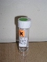
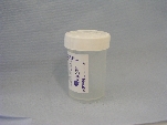
Specimen container
Sterile 30 ml universal
Make sure lid is firmly secured
Transport to the laboratory If transport is delayed then refrigerate sample. Delays of over 48 hours are undesirable
Turnaround 5 working days
Preparation method used by the laboratory Cytospin, Papanicolaou and Romanovsky May Grunwald Giemsa.
Factors known to significantly affect the performance of the examination or the interpretation of results The nature of the abnormality being investigated should be clear from the clinical information included with the specimen so that the laboratory can perform the appropriate preparations.

Imaging guidance may be required to successfully target some lesions.

A negative result of an exfoliative cytology sample is not sufficient evidence to exclude significant disease. Discussion of cases at multi-disciplinary team meetings and submission of further samples, if deemed clinically appropriate, should thus be a routine part of the diagnostic pathway. Cytology results should be correlated with histology findings. Audit against final outcomes (which may be clinical) should be performed.
Additional information Please ensure that all request forms and specimen pots are clearly labelled with patient details and relevant clinical information.
Use a Non-Gynae request form/ICE request form


FINE NEEDLE ASPIRATION CYTOLOGY

Investigation Non-Gynae Cytology
Inform lab before sending No (unless very urgent: extension 5623)
Specimen type Fine needle aspiration is widely accepted as a first line of investigation in the diagnosis of focal mass lesions.
Material obtained by aspiration may be directly spread onto a slide or placed in collection fluid (CytoLyt). Ideally, a combination of direct slide and material washed into collection fluid should be submitted using the following procedure:

1. Prepare 2-4 air dried slides (blood film type preparation).


Rapid drying of the material is important in preserving the cellular detail.


2. ALL slides prepared must be labelled using a pencil and include
• Patient's First name
• surname
• D.O.B. or Unit number (e.g. RQ6/NHS) and Site of FNA
3. Place these completely dried and labelled slides in a slide carrier.
4. Wash out the rest of the specimen from the needle and syringe in to CytoLyt.
5. Send both slides and fluid to Non-Gynae Cytology.
Maximum Volume Required 20 mls
Specimen container
30 ml universal containing 10 mls of Cytolyt fluid.

These are obtained from Non-Gynae Cytology ext 5623.

All bottles have an expiry date printed on them.

Make sure lid is firmly secured

Transport to the laboratory If transport is delayed specimen will keep for about 1 month.
Turnaround 5 - 10 working days
Preparation method used by the laboratory Direct smears, May Grunwald Giemsa.
Liquid based cytology, Papanicolaou.
Factors known to significantly affect the performance of the examination or the interpretation of results The nature of the abnormality being investigated should be clear from the clinical information included with the specimen so that the laboratory can perform the appropriate preparations.

Accurate targeting of the lesion is essential for a reliable diagnosis. Depending on the size and location of the lesion, targeting may be by palpation or imaging. Unless easily palpable, the use of ultrasound for targeting significantly increase the diagnostic yield.

FNAs should not be taken by unsupervised staff who have no training in the technique, staff who do not take such aspirates regularly, or who are unaware of the risks and complications.

Needles:
Needles should be 23 gauge or less. It is important to use smaller needles as they cause less bleeding, the risk, albeit rare, of tumour seeding is considerably reduced, and it is less painful for the patient. Thicker needles (18G or wider) carry an ever-increasing risk of complications including significant haemorrhage. Needles should have a long bevel giving a relatively large circumference at their cutting edge. Longer fine needles may be required for deeply sited lesions targeted by image guidance. Without a stylet a long needle may be uncontrollable and dangerously flexible.

Staff making the preparations must be suitably trained to ensure the sample is prepared appropriately.

A monolayer of cells is required therefore under-spreading of the sample can significantly hamper analyses.
Overspreading of the sample can result in the destruction of the cellular structure and therefore significantly hamper analyses.

Spread samples should be air-dried rapidly and completely to prevent the cells from lysing. The most effective method is using a hairdryer on a cool setting. If a slide is placed into a slide carrier whilst wet then all of the slides in that carrier will be adversely affected by the moisture created.
Additional information Where there is a clinical suspicion of an infectious disease (Tuberculosis, HIV, Hepatitis B, etc), direct smears should not be prepared. In these cases all of the material should be washed directly into CytoLyt.

Breast - Assessment of oestrogen and progesterone hormone receptors by immunocytochemistry and HER-2 status by immunocytochemistry or FISH is required for all invasive breast carcinomas. These assessments may be performed on either cytological or histological material, providing sufficient is available. Whilst these investigations will often be easier to perform on a core biopsy, there are clinical settings where this is not in the best interests of the patient. In these instances, laboratories will undertake these investigations on cytology specimens.

Special precautions:
There are specific contraindications and potential risks to deep site aspirates.
• Anticoagulant therapy and intrinsic bleeding problems increase the risk of bruising and haemorrhage.
• Intractable cough and poor respiratory function are absolute contraindications to transthoracic FNA.
• FNA of carotid body tumours may cause catecholamine release with the potential risk of hypertensive crisis.

Please ensure that all request forms and specimen pots are clearly labelled with patient details and relevant clinical information. Use a Non-Gynae request form/ICE request form


Health and safety

All fresh body fluids may harbour unexpected biohazards, such as mycobacteria or hepatitis B, C or human immunodeficiency virus (HIV), it is essential that appropriate precautions are taken to prevent infection of staff and patients.

Safe Disposal of Materials used in sample collection

• All sharps must be used and disposed of safely in line with the Trust Safe use and disposal of sharps policy.
• In known high-risk patients FNA needles should not be separated from the syringe at the time of disposal in to the sharps container.
• Other materials used in sample collection should be cleaned as per local protocols/manufacturers instructions or disposed of as clinical waste using the appropriate yellow clinical waste bags.

Sequential Testing of samples

As well as the routine preparation methods employed by the laboratory sequential testing may be required for some samples to yield further information. Sequential testing may take the form of additional preparations to allow more material to be analysed, fixation of the sample in formalin for the preparation of a cell block, immunocytochemistry and molecular pathology techniques, and special staining techniques to demonstrate other components of the sample not seen with routine Papanicolaou and Romanovsky/Giemsa stains.

Immunocytochemistry:
Immunocytochemistry should be used to investigate possible malignant disease and to establish differentiation as appropriate. In general, it is particularly important to distinguish lymphoma, melanoma, carcinoma, mesothelioma and sarcoma as this has significant implications for management. Immunocytochemistry for various tissue specific antigens and cytokeratins may be used to suggest or support a primary site in metastatic disease.

Flow cytometry is the method of choice for confirming and typing lymphoma in fluid samples. The appropriate transport medium should be agreed locally. Rapid transit is required.

Complaints Procedure

Complaints can either be made verbally or formally in writing. If you wish to raise a concern or complaint regarding the service, please contact Jane Harrison-Williams, 0151 706 4509, jane.harison-williams@LiverpoolFT.nhs.uk Cellular Pathology Quality Practitioner

References:

Tissue Pathways for exfoliative cytology and fine needle aspiration cytology (G086). The Royal College of Pathologists, v1. January 2010
Dr Karin Denton, North Bristol NHS Trust (Lead author); Dr Thomas Giles, Royal Liverpool and Broadgreen Hospitals NHS Trust; Dr Peter Smith, Royal Liverpool and Broadgreen Hospitals NHS Trust; Dr A Ashish Chandra, Guys and St Thomas’ NHS Trust; Dr Mina Desai, Manchester Royal Infirmary.

The BSCC Code of Practice – exfoliative cytopathology (excluding gynaecological cytopathology). Cytopathology 2009, 20, 211-223. A. Chandra, P. Cross, K. Denton, T. Giles, D. Hemming, C. Payne, A. Wilson and P. Wilson.

BSCC code of Practice – fine needle aspiration cytology. Cytopathology 2009, 20, 283-296. G. Kocjan, A. Chandra, P. Cross, K. Denton, T. Giles, A. Herbert, P. Smith, D. Remedios and P. Wilson.