Immunocytochemistry
Pathology

Contacts: Sharon Forrest
Immunocytochemistry & Molecular Pathology Services Manager
Tel: 0151 706 4485
Email: Sharon.Forrest@rlbuht.nhs.uk

Tracey Galvin
Immunocytochemistry Team Leader
Tel: 0151 706 4485
Email: Tracey.Galvin@rlbuht.nhs.uk


Location of Laboratory: The laboratory is situated on the 3rd Floor CSSB.

Services offered by the laboratory: Immunocytochemistry (ICC) is the in-situ detection and demonstration of cellular constituents using specific antigen-antibody reactions. The technique has evolved to become an integral diagnostic aid in the modern Cellular Pathology Department. Both histological and cytological material is tested where appropriate to refine the diagnosis, determine prognostic indicators or to assess specific indicators of tumour response.

The Department's Immunocytochemistry laboratory is fully automated, and is able to offer a range of approximately 200 different primary antibodies to provide immunoperoxidase, immunofluorescence (IMF) and in situ hybridization (ISH) slide-based methodologies to service users. The laboratory also undertakes HER2 testing on breast tumour samples by immunocytochemistry (using the 4B5 clone) and fluorescence in situ hybridisation (FISH) and offers these as a regional service.

Laboratory opening hours: Monday to Friday 07:00-19:00.

Sample types: immunoperoxidase and in situ hybridisation techniques require formalin fixed paraffin embedded (FFPE) tissue sections cut at a thickness of 4microns and mounted onto APES coated slides.

IMF techniques require fresh frozen tissue sections; samples should be transported to the lab in normal saline solution or in Michel’s medium.

Sample volumes: section thickness for immunoperoxidase and in situ hybridisation (ISH) techniques should be 4microns.

Cryostat sections for IMF protocols should be cut at a thickness of 5microns.

Special precautions: sections for routine immunoperoxidase stains should be heated onto APES slides overnight; a heating temperature range of 37-50°C is acceptable for routine ICC staining protocols. Alternatively, sections for routine immunoperoxidase stains can be heated for 30minutes at 60°C.

Sections for HER2 staining should be cut onto APES slides and heated overnight at a temperature of 37°C. The Superfrost Plus sialinized slide type is the recommended option when preparing slides for HER2 ICC.

Sections for ISH should be cut onto APES slides and heated overnight; a temperature range of 37- 50°C is acceptable. The Superfrost Plus sialinized slide type is the recommended option when preparing slides for in situ hybridisation (FISH and CISH) techniques.

Fresh tissue for IMF procedures should be transported to the lab in a timely manner in saline solution or Michel’s medium.

Turnaround times (TATs): 10 working days for routine immunocytochemistry stains. TATs for HER2 and ISH analyses are 14 working days.

Instructions for completion of request forms: refer to the Liverpool Clinical Laboratories Minimum Dataset Policy (MDS) for Laboratory Investigations for guidance. Link

Instructions for transportation of samples: slides for routine immuoperoxidase stains, HER2 or in situ hybridisation techniques should be labelled with the sample’s unique laboratory number and patient surname and transported to the laboratory in sealed slide mailer boxes.

Fresh tissue for IMF analysis should be transported to the lab in saline solution or in Michel’s medium and the specimen pot labelled in accordance with the guidelines outlined in the MDS Policy for Histology samples.

Patient consent: not applicable.

Lab criteria for accepting/rejecting samples: criteria for sample acceptance or rejection is outlined in the MDS Policy; please see this Policy document for advice. Link

Factors known to significantly affect performance of examination or interpretation of results: prompt, adequate fixation of samples is imperative for immunocytochemical and in situ hybridisation analyses. Tissue should be placed into an adequate volume of fixative (10X the volume of the sample) as soon as possible after excision to protect against the deleterious effects of cold ischemia on ICC and ISH staining.

Sections for routine ICC and HER2 analyses should be cut fresh as immunoreactivity of certain antigens, i.e. ER, PGR and HER2 is subject to oxidation and has been shown to decline with section age.

It is recommended that sections for HER2, HERFISH and CISH analyses are mounted onto Superfrost Plus sialinized slides. Artefacts associated with the use of other adhesive slide types can impede interpretation.

Availability of clinical advice when ordering examinations and on interpretation of examination results: technical advice can be obtained by contacting the Immunocytochemistry and Molecular Services Manager or Immunocytochemistry Service Lead by e-mail (see above for contact details) or on 0151 706 4485.

Clinical advice on ICC examination results can be obtained by contacting the relevant member of the Consultant Pathologist team.

Protection of personal information: the laboratory complies with the mandates set out in the Data Protection Act and Caldicott regulations. Trust and local policies are also in place to assure the protection of personal information.

Complaints procedure: if you wish to raise any concerns regarding the ICC service, please contact the Immunocytochemistry and Molecular Services Manager, Sharon Forrest, on 0151 706 4485 or by e-mail at sharon.forrest@LiverpoolFT.nhs.uk. Alternatively concerns can be raised by contacting the cellular Pathology Clinical Director Dr Vijay Sharma via email vijay.sharma@LiverpoolFT.nhs.uk or the Quality Practitioner Jane Harrison-Williams 0151 706 2000 ext 11093 or email jane.harrison-williams@LiverpoolFT.nhs.uk.
To view the full document click here.


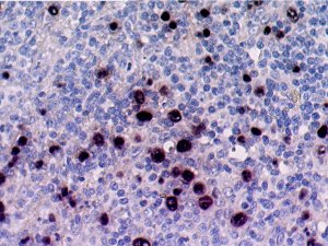
Epstein-Barr virus-encoded RNA (EBER) demonstrated by in-situ hybridisation in a case of post-transplant lymphoproliferative disease.

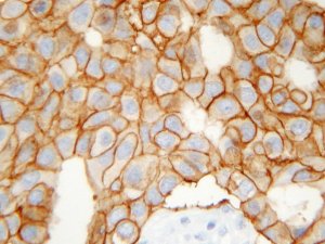
Over expression of HER2, a member of the erbB epithelial growth factor receptor family in a breast carcinoma case.

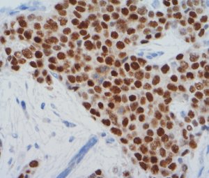
An oestrogen receptor positive breast carcinoma.

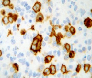
CD30 positive Reed-Sternberg cells in a case of Hodgkin's disease.

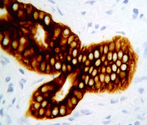
A normal duct, the epithelial cells of which have been stained using an antibody to cytokeratins (an intermediate filament protein).

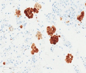
Groups of mesothelioma cells in a non-gynae cytology EBUS specimen which have been stained using an antibody to calretinin (which is expressed in both normal and neoplastic mesothelial cells and is a useful marker for the identification of malignant mesothelioma of the epithelial type).